Together, we can positively impact patients living with vitiligo
Vitiligo is a dermatological disease that's more than what you see on the surface. Vitiligo is a chronic, immune-mediated pigmentary skin disorder that may significantly impact quality of life and pose considerable burden on patients and caregiver.
Learn more about vitiligo and its impact below:

Disease Education
The better we understand vitiligo, the better prepared we will all be in helping patients gain control over this condition.
LIVING WITH VITILIGO
Vitiligo Is a Chronic Autoimmune Skin Disease
Non-scaly macules and patches devoid of pigment1 with distinct margins due to melanocytic destruction.2
Presents anywhere on the body3-5 but most commonly on the face (87.0%), acral areas* (76.3%), and extremities (59.7%).6
Nonsegmental vitiligo (NSV) is the most common form of vitiligo (80%–90% of cases),3 characterized by symmetrical white patches on both sides of the body.1
NSV has an unpredictable disease course—even stable disease may become active without adequate control of immune-mediated processes.1
EPIDEMIOLOGY
Most common depigmenting skin disorder.1,7
Affects 0.76%–1.11% of adults in the US.8
Regardless of sex, racial, or ethnic groups.9-11
Develops at any age, but is more common in younger people,1,10,11 with 70%–80% of cases occurring before 30 years of age.11
PROGNOSIS
Disease prognosis may depend on:
Age of onset12
Extent of disease12
Disease type (segmental vs non-segmental)13,14
Lesion location15,16
*Acral distribution of skin lesions involves the distal aspects of the head (ears, nose) and the extremities (hands, fingers, feet, toes).
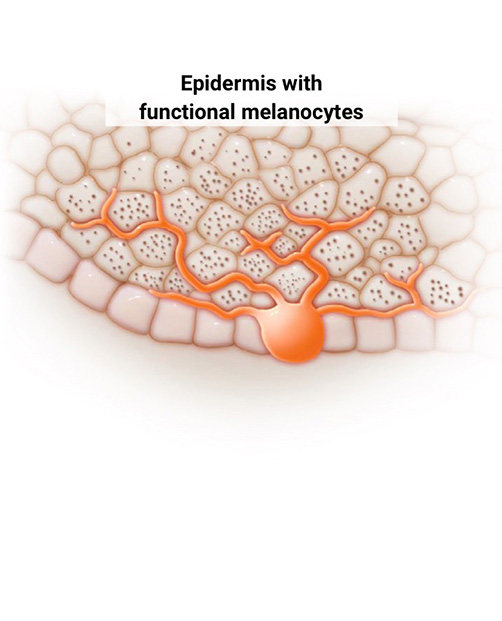
THE PIGMENTATION OF HEALTHY SKIN
Melanocytes Play a Key Role in the Pigmentation of Healthy Skin17,18
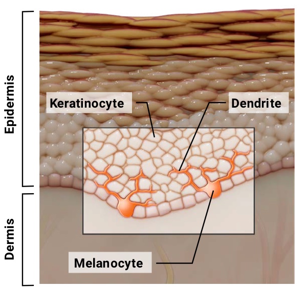
Melanocytes are found in the base layer of the epidermis. Each melanocyte extends dendrites into the surrounding epidermis to interact with keratinocytes.
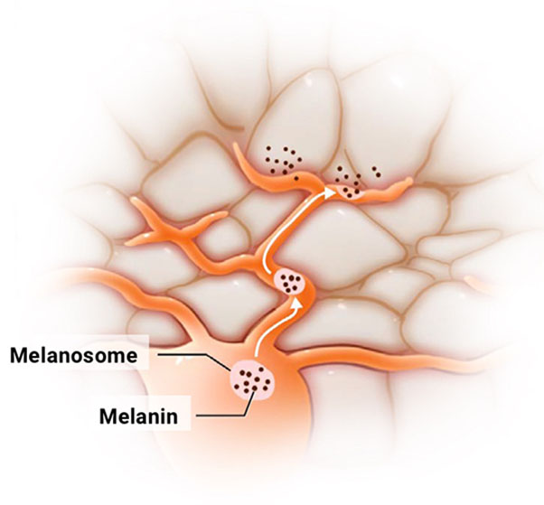
During melanogenesis, melanocytes produce melanin in melanosomes, then use their dendrites to transfer the melanosomes to keratinocytes.
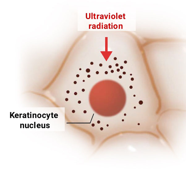
Once inside a keratinocyte, the melanosomes distribute uniformly to ensure even pigmentation of the skin and create a shield to protect the nucleus from ultraviolet radiation.
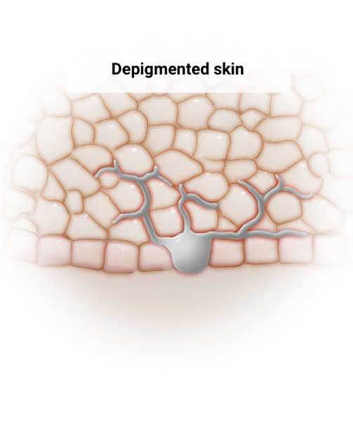
THE IMMUNE SYSTEM’S ROLE IN THE PATHOGENESIS OF VITILIGO
Vitiligo Is a Depigmenting Skin Disease in Which the Immune System Attacks Melanocytes19,20
Treatment of vitiligo involves controlling inflammation and stabilizing the disease, which facilitates melanocyte-induced melanin production and repigmentation.21,22
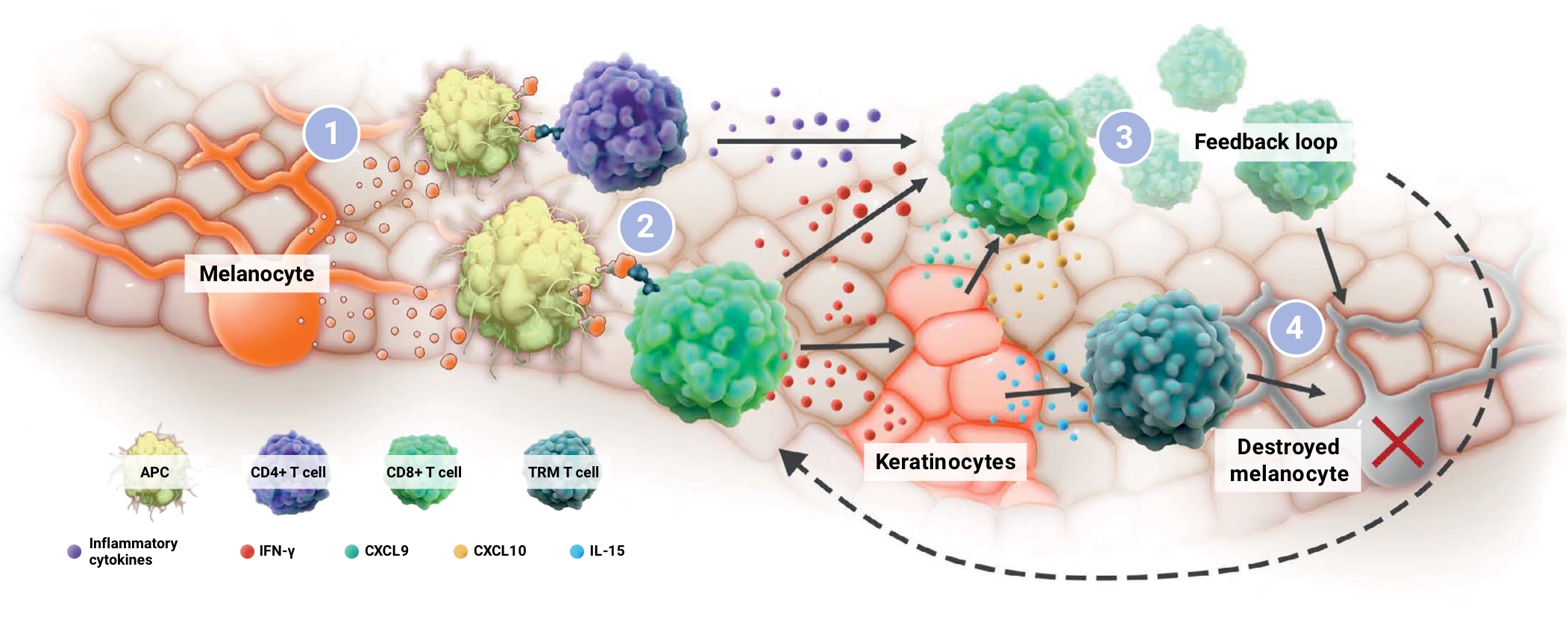
APC, antigen-presenting cell; CD, cluster of differentiation; CXCL, chemokine ligand; IFN, interferon; IL, interleukin; JAK, Janus kinase; STAT, signal transducer and activator of transcription; TRM, tissue-resident memory. Adapted from Qi F et al. Front Immunol. 2021;12:790125.
MELANOCYTE SOURCES FOR REPIGMENTATION
Repigmentation Requires Time for Melanocyte Proliferation, Migration, and Differentiation34,35
Repigmentation is a gradual process that takes time.36
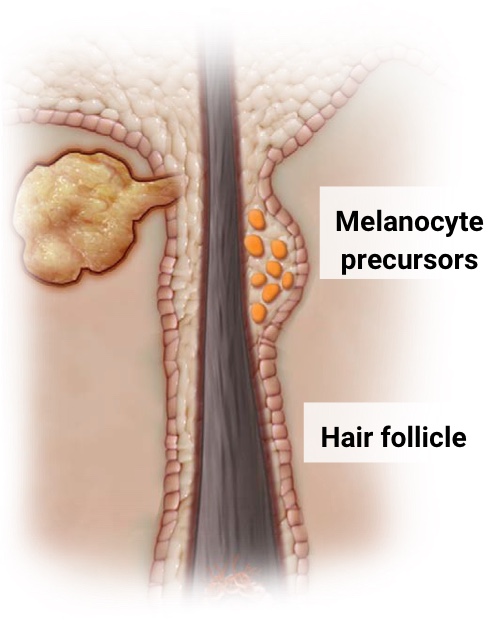
Melanocyte precursors in the immune privileged hair follicle are protected from T-cell attack.34,35
By regulating the aberrant immune response, activated melanocyte precursors proliferate and migrate to the depigmented skin.34
The epidermis at the edge of lesions contains functional melanocytes, representing a secondary source for repigmentation.34,36
BEYOND THE SKIN
Vitiligo Explained: Understanding the Mechanism of Disease
References: 1. Ezzedine K et al. Lancet. 2015;386:74-84. 2. Rashighi M et al. Dermatol Clin. 2017;35:257-265. 3. Al-smadi K et al. Cosmetics. 2023;10:84. 4. Mazzei Weiss ME. Cutis. 2020;105:189-190. 5. Joge RR et al. Cureus. 2022;14:e29307. 6. Speeckaert R et al. J Eur Acad Dermatol Venereol. 2014;28:755-762. 7. Bergqvist C et al. Dermatology. 2020;236:571-592. 8. Gandhi K et al. JAMA Dermatol. 2021;158:1-96. 9. Alikhan A et al. J Am Acad Dermatol. 2011;65:473-491. 10. Alkhateeb A et al. Pigment Cell Res. 2003;16:208-214. 11. Sehgal VN et al. Indian J Dermatol Venereol Leprol. 2007;73:149-156. 12. Ahmed JN et al. Vitiligo. StatPearls [Internet]; 2023. 13. Delbaere L et al. Dermatol Rev. 2022;289-307. 14. Lin F et al. Sci Rep. 2021;11:18298. 15. Njoo MD et al. Arch Dermatol. 1998;134:1532-1540. 16. Taieb A et al. Br J Dermatol. 2013;168:5-19. 17. Maranduca MA et al. Oncol Lett. 2019;17:4183-4187. 18. Bento-Lopes L et al. Int J Mol Sci. 2023;24:11289. 19. Ezzedine K et al. Pigment Cell Melanoma Res. 2012;25:E1-13. 20. Dell’anna ML et al. Pigment Cell Res. 2006;19:406-411. 21. Cunningham KN et al. Am J Clin Dermatol. 2023;24:165-186. 22. Li W et al. Curr Issues Mol Biol. 2025;47:191. 23. Post NF et al. Pigment Cell Melanoma Res. 2023;36:348-354. 24. Wang J et al. Redox Rep. 2022;27:193-199. 25. Harris JE et al. J Invest Dermatol. 2012;132:1869-1876. 26. van den Boorn JG et al. J Invest Dermatol. 2009;129:2220-2232. 27. Rashighi M et al. Sci Transl Med. 2014;6:223ra223. 28. Ivashkiv LB. Nat Rev Immunol. 2018;18:545-558. 29. Harris JE. Immunol Rev. 2016;269:11-25. 30. Hlača N et al. Biomedicines. 2022;10:1639. 31. Custurone P et al. Int J Mol Sci. 2021;22:11429. 32. Qi F et al. Front Immunol. 2021;12:790125. 33. Su X et al. Front Immunol. 2025;16:1639732. 34. Birlea SA et al. Dermatol Clin. 2017;35:205-218. 35. Falabella R. Indian J Dermatol. 2009;54:313-318. 36. Passeron T. Dermatol Clin. 2017;35:163-170.